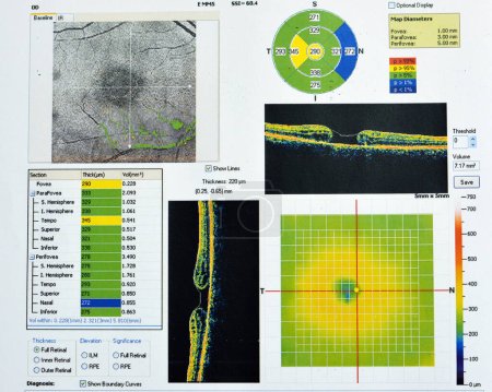
OCT of the eye reveals faint epimacular membra ... 

Media-ID: B:657057702
Nutzungsrecht:
Kommerzielle und redaktionelle Nutzung
OCT of the eye reveals faint epimacular membrane and full thickness macular hole involving the fovea, surrounding diffuse macular oedema showing few cystoid changes for follow up, selective focus
| Vorschau |
Varianten
Mediainfos
|
Dieses Bild mit unserem Kundenkonto ab $1.25 herunterladen!
|
||||
| Standardlizenz: JPG | ||||
| Format | Bildgröße | Downloads | ||
|
Print XXL 15 MP |
4800x3840 Pixel 40.64x32.51 cm (300 dpi) |
1 | ||
| Standardlizenz: JPG | ||||
| Format | Bildgröße | Netto | Brutto | Preis |
|
Web S 0.5 MP |
500x400 Pixel 16.93x13.55 cm (75 dpi) |
$5.15 | $5.50 | |
|
Print M 2 MP |
1000x800 Pixel 8.47x6.77 cm (300 dpi) |
$9.11 | $9.74 | |
|
Print XL 8 MP |
2000x1600 Pixel 16.93x13.55 cm (300 dpi) |
$17.03 | $18.22 | |
|
Print XXL 15 MP |
4800x3840 Pixel 40.64x32.51 cm (300 dpi) |
$20.99 | $22.45 | |
| Merchandisinglizenz: JPG | ||||
| Format | Bildgröße | Netto | Brutto | Preis |
|
Print XXL 15 MP |
4800x3840 Pixel 40.64x32.51 cm (300 dpi) |
$105.47 | $112.85 | |
| Media-ID: | B:657057702 |
| Aufrufe: | 1 |
| Beschreibung: | OCT of the eye reveals faint epimacular membrane and full thickness macular hole involving the fovea, surrounding diffuse macular oedema showing few cystoid changes for follow up, selective focus |
Nutzungslizenz
| Nutzungsrecht: | Kommerzielle und redaktionelle Nutzung |
Userinfos
| Hinzugefügt von: | Tamer_Soliman |
| Weitere Medien von Tamer_Soliman |
Bewertung
| Bewertung: |
|
Suchbegriffe
| Keywords: |


