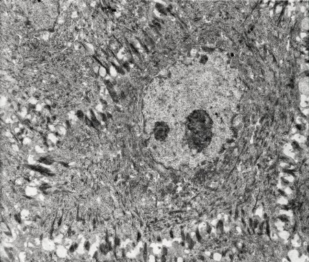
Epidermis. Electron microscope micrograph show ... 

Media-ID: B:713926574
Nutzungsrecht:
Kommerzielle und redaktionelle Nutzung
Epidermis. Electron microscope micrograph showing a keratinocyte of spinous layer. The epithelial cell has a polygonal shape, central nucleus with nucleolus, cytoplasm full of keratin filament bundles, and numerous dark desmosomes crossing the interc
| Vorschau |
Varianten
Mediainfos
|
Dieses Bild mit unserem Kundenkonto ab $1.25 herunterladen!
|
||||
| Standardlizenz: JPG | ||||
| Format | Bildgröße | Downloads | ||
|
Print XXL 15 MP |
3984x3384 Pixel 33.73x28.65 cm (300 dpi) |
1 | ||
| Standardlizenz: JPG | ||||
| Format | Bildgröße | Netto | Brutto | Preis |
|
Web S 0.5 MP |
500x425 Pixel 16.93x14.39 cm (75 dpi) |
$5.15 | $5.50 | |
|
Print M 2 MP |
1000x849 Pixel 8.47x7.19 cm (300 dpi) |
$9.11 | $9.74 | |
|
Print XL 8 MP |
2000x1699 Pixel 16.93x14.38 cm (300 dpi) |
$17.03 | $18.22 | |
|
Print XXL 15 MP |
3984x3384 Pixel 33.73x28.65 cm (300 dpi) |
$20.99 | $22.45 | |
| Merchandisinglizenz: JPG | ||||
| Format | Bildgröße | Netto | Brutto | Preis |
|
Print XXL 15 MP |
3984x3384 Pixel 33.73x28.65 cm (300 dpi) |
$105.47 | $112.85 | |
| Media-ID: | B:713926574 |
| Aufrufe: | 1 |
| Beschreibung: | Epidermis. Electron microscope micrograph showing a keratinocyte of spinous layer. The epithelial cell has a polygonal shape, central nucleus with nucleolus, cytoplasm full of keratin filament bundles, and numerous dark desmosomes crossing the interc |
Nutzungslizenz
| Nutzungsrecht: | Kommerzielle und redaktionelle Nutzung |
Userinfos
| Hinzugefügt von: | [email protected] |
| Weitere Medien von [email protected] |
Bewertung
| Bewertung: |
|
Suchbegriffe
| Keywords: |


