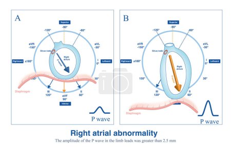
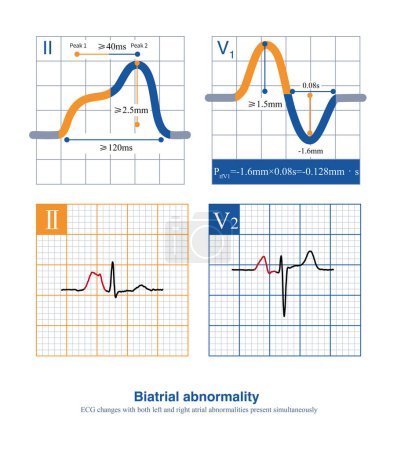
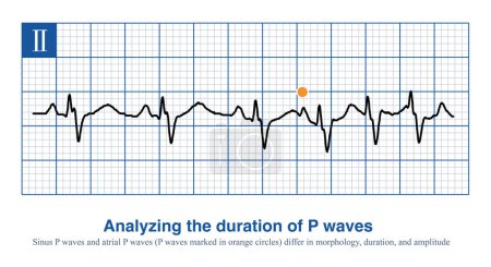
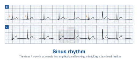
5 Bilder zum Thema "p wave amplitude" bei ClipDealer

Medien sortieren nach     Gehe zu Seite:    
« Vorherige  1  Nächste »
In patients with emphysema, the anatomical position of the heart is more vertical, the depolarization potential of the right atrium from top to bottom increases, and the P wave of the ECG increases.
When the duration of the P wave exceeds 120 ms, the amplitude of the limb leads exceeds 2.5 mm, and the amplitude of the thoracic leads exceeds 1.5 mm, it is interpreted as a biatrial abnormality.
When intraatrial excitation is transmitted from one atrium to the other, a wider duration of P waves are generated, such as sinus rhythm, lateral  left atrial focal, etc.
Sometimes, the P wave amplitude of sinus rhythm is so low that it is close to an isopotential and can easily be mistaken for a borderline rhythm.
Male, 10 years old, was clinically diagnosed with tetralogy of Fallot. ECG shows an elevated P wave amplitude in thoracic leads, suggesting right atrial abnormality.

« Vorherige  1  Nächste »